장비성능
※ 신청 전 담당자에게 문의 바랍니다. - 담당자 : 전진혁 (042-865-3490)
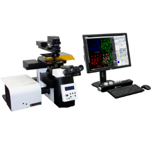
☞ 고해상도 이미징 및 표면 분석을 위한 공초점 현미경
☞ 물리적 절단 없이 시료에서 발광되는 형광 빛을 이용하여, 광학 절단된 단면 이미지를 만들 수 있고 이를 조합하여 3D을 형상화할 수 있음
Laser Module
Microscope Module
- Adaptable sample stage: glass slides, well plates, Petri-dishes
- XY stage: Motorized or manual stage, Stroke 115 mm x75 mm ; various travel range customizable
- Z-drive: Motorized stage - 15mm travel range / 250nm step size min.
PZT stage (single objective lens) - 400µm travel range / 1nm step size
- Accessories: Digital Cameras – sCMOS, high sensitivity CMOS, cooled-CCD, etc.
Detector Module
- Detection range: 400-750nm or NIR detection is customizable
- Number of detector: Up to four PMTs with each emission filter
- Sensor: Standard : Highly sensitive PMT
Low light model : Ultra-highly sensitive GaAsP PMT
Scanner Module
- Scanners: Resonant scanner and galvanometer mirror
- Scan resolution: 128x128 ~ 2048x2048 selectable
- Scan speed: 30fps at 512 x 512 pixels (Bi-scan), 15fps at 512 x 512 pixels (Uni-scan)
- Scan zoom: 0.7x~3x continuously variable
- Scan field: Square 12.5mm divided by objective lens magnification (Field number 18)
- Scan mode: xy, xyz, xt, xyt, xyzt
- Pinhole: Motorized switching pinhole