Center for Scientific Instrumentation
Center for Scientific Instrumentation Division of R&D Equipment Industry > Major Research Activities Home
Center for Scientific Instrumentation builds and operates cutting-edge research equipment, and develops new analytical methods to conduct creative research support and joint research with domestic and foreign universities, research institutes and companies. In addition, we develop scientific research equipments to secure the core technologies and foster the domestic research equipment industry.
Main Research Field
Mass Spectrometry and Advanced Instrumentation Researcg
- Development of 3-Dimensional Molecular Imaging Mass Spectrometry
- Development of key component technologies of mass spectrometry and various ion beams and its application
Electro-Mgnetic Properties measurement System (EMPS)
- Development of electro-magnetic properties measurement system and fundamental technology
- Development of property analysis platform and its application
Multi-modal optical imaging system instrumentation
- Development of multi-modal (optical coherence, multi photon, thermal reflectance, fluorescence) microscopy and its application
- Development of high speed confocal fluorescence microscope
1st Domestically development of Transmission Electron Microscope (TEM)
- Development of key component technologies of low-end TEM
- Development of key component technologies of Cs-corrected TEM
Development of 3D organ models (organ-on-a-chip and organoids) and its analysis systems
- Development of analytical models and instruments for 3D organ models
- Development of analytical software based on artificial intelligence (AI) for 3D organ models
Development of Thermal Imaging Microscope
- Development of high resolution thermo-reflectance microscope
- Development of thermal analysis technology for microelectronic devices
Representative Research Cases
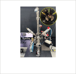
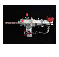
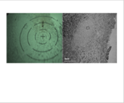
The first development of time-of-flight secondary ion mass spectrometry
- Developed gas cluster ion beam, the core technology of time-of-flight secondary ion mass spectrometry, for the first time in Korea. We are developing a time-of-flight secondary ion mass spectrometer capable of analyzing organic molecules and biological samples using cluster ion beams. Conduct research and commercialization tasks for local production of mass spectrometers using developed key element technologies.
-
 3D molecular imaging mass spectrometer
-
 Gas cluster ion beam
-
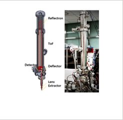 Time-of-Flight mass
spectrometer
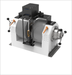
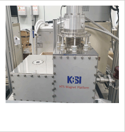
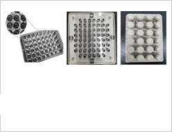
Development and commercialization of Electro-Magnetic Properties measurement System (EMPS) using an electromagnet
- Development and commercialization of Electro-Magnetic Properties measurement System (EMPS) measuring hall coefficient as well as magnetic characteristics under variable magnetic field was successfully carried out. It has higher magnetic field efficiency than commercial equipment and will be applied to the development of new material and components using an advanced analysis technology.
-
 Electro-magnetic properties
measurement using electromagnet -
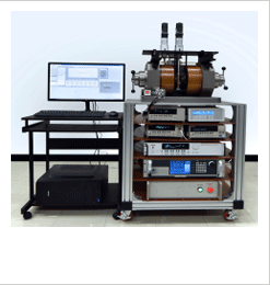 Hall coefficient measurement
system using electromagnet -
 Conduction-cooled magnetic
field generation platform
Development of a multi-mode optical microscope capable of live cell imaging
- By combining optical coherence, confocal fluorescence, and nonlinear (second harmonic, third harmonic) imaging modes that enable optical sectioning of thick samples such as 3D samples (organoids, spheroids), the co-registered imaging of three-dimensional structure and specific molecule in the bio sampels is possible. In order to reduce photobleaching/photodamage that often occurs in live cell fluorescence imaging, we proposed a method to find regions of interest by optical coherence imaging and enable high-resolution fluorescence imaging.
-
 Multi-mode optical
microscope -
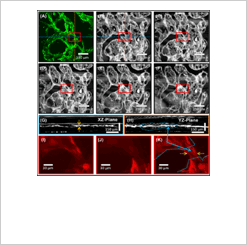 Finding regions of interest
in 3D vascular organoids -
 Cancer cell spheroid imaging
cultured for 5 days
The first development of low-end transmission electron microscope
- We developed 1st testbed of 30kV transmission electron microscope (TEM) which have FEG system. This instrument have advantages of minimizing the beam damages and enhancement of image contrast. Therefore, it is expected that low-end TEM could be optimized and useful tool for imaging of light element or biological samples.
-
 1st testbed of TEM
-
 Image & diffraction pattern
of Au standard sample -
 Images of mouse brain hippocampus
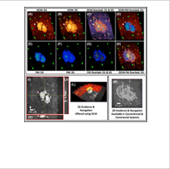
Development of a fully integrated system with 3D organ model, devices, and analysis software
- We develop a biochip-based optical device integrated with a 3D biological model, featuring a cutting-edge AI-based 3D image analysis platform. For implementation, we design relevant devices, implement processing methods, and establish automated analysis systems. The use of high-throughput 3D organ models based on well plates, coupled with AI-based image analysis, enables the rapid and precise evaluation of newly developed drugs. This approach shows great promise in advancing efficacy assessments in drug development and offering a valuable alternative to traditional animal models.
-
 Well plate-based 3D organ
analysis platfrom -
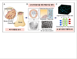 Bone-mimetic osteoprosis model
and AI-based AI algorithm -
 Automation system for
multi-organ analysis
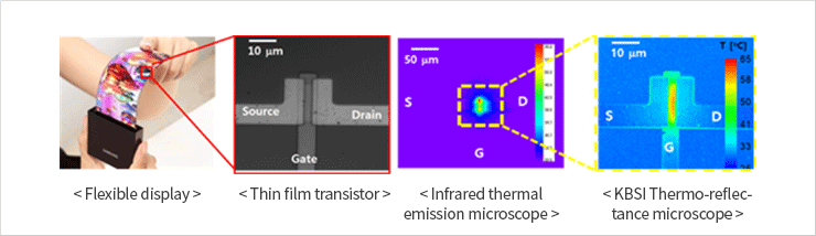
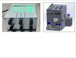
Development of the world’s best performance thermal microscope
- We have developed a thermo-reflectance microscopy technique capable of visualizing temperature distributions of micro-electronic devices such as semiconductors, displays, sensors, and light-emitting diodes (LEDs). This technique offers spatial resolution of 0.3 μm and temporal resolution of 1.0 μs, and allowing for imaging surface and sub-surface heat distribution of samples.