Representative Research Publications
Development of new laser spectroscopic technique distinguishing between mirror-imaged structures of 2018 > Representative Research Publications > Research Results Home
Development of new laser spectroscopic technique distinguishing between mirror-imaged structures of enantiomers
- Journal of Physical Chemistry Letters / 2018 December
- Taegon Lee(1st author), Hanju Rhee(Corresponding author)
Study Summary
Chiral molecule is not superimposable with its mirror image like human (left and right) hands. Note that a pair of non-superimposable mirror-imaged molecules is called enantiomer or mirror (or stereo) isomer. These chiral molecules often have different biochemical activities and their distinct structure plays a critical role in our body. For instance, one form of enantiomeric chiral drugs has a good efficacy but the other mirror-imaged form is ineffective or even harmful to life. To understand their function and underlying mechanism in life, an incisive tool for characterizing stereochemical structure of chiral molecule at the microscopic scale is needed.
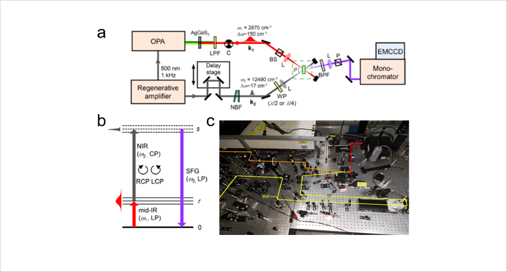
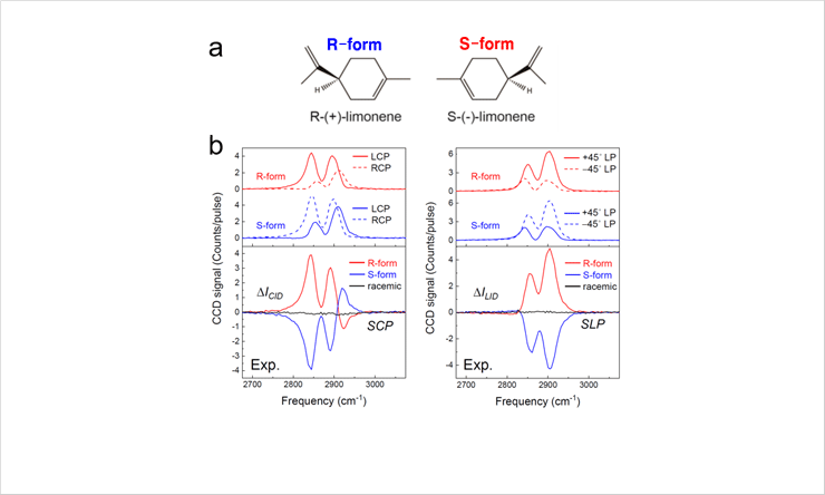
We developed a new second-order nonlinear spectroscopic technique, called femtosecond chiral vibrational sum-frequency generation (SFG), sensitive to stereo-specific vibrational structure of chiral molecule. In contrast to the conventional methods employing only a single IR or visible laser beam, we combined two laser beams (femtosecond mid-IR and picosecond visible lasers) to obtain the distinguishable chiral vibrational SFG spectra of R- and S-limonene, which are a pair of enantiomers. By properly controlling the polarization states of the involved laser fields, we could selectively isolate the chiral component from the generated nonlinear signals to dramatically enhance the chiral sensitivity as well as signal-to-noise ratio.
 Fig. 1. Experimental layout (a), energy-level diagram (b) and photographic image (c) for chiral vibrational SFG measurement
 Fig. 2. Chemical structures of R- and S-limonene (a) and their mirror-imaged chiral vibrational SFG spectra measured with the experimental setup in Fig. 1
The chiral vibrational SFG technique can be used to study ultrafast dynamics involving chiral structural changes such as protein folding/unfolding, DNA-drug binding and photoisomerization processes. Furthermore, it can be extended to stereochemical imaging technique for elucidating pharmacological function and mechanism of chiral drugs in living system. We anticipate that the present technique will be of critical use in developing ultrafast transient chiroptical spectroscopy and chiral vibrational SFG microscope in the near future.


